Lecture 10. Proteins: Supersecondary and tertiary structure
Wednesday 28 September 2016
Building up tertiary structure from secondary structure elements: Motifs, supersecondary structure, and domains. Topology diagrams. Methods for protein structure determination. Structure is conserved more than sequence. Classification of tertiary structures. Quaternary structure and symmetry.
Reading: VVP4e - Ch.6, pp.127-141.
Summary
Proceeding from secondary structure "elements", we can describe how these elements are linked in a particular protein, and the order of β strands in a β sheet structure. This is called protein topology, and can be represented by means of a topology diagram (see figure at below). The topology diagram helps us see how the three-dimensional structure of a long polypeptide chain - i.e. protein tertiary structure - can be built up from its secondary structure elements. Topological representation, although abstract, reveals patterns commonly repeated and reiterated throughout structural biology. The recognition of motifs or supersecondary structures as simple "compounds" of secondary structure that bridge the hierarchical gap between secondary and tertiary protein structure can be seen in this light. Given the many thousands of protein structures that have been obtained by the methods of X-ray crystallography and multidimensional NMR, a number of general principles of protein tertiary structure seem well-established.
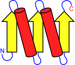
Left: Example of a topology diagram for a polypeptide chain. The parts of the chain that adopt β-strand conformation are shown as yellow arrows, α-helices are shown as red cylinders. These secondary structure elements are connected by the blue loops
Another level of the protein structure hierarchy, quaternary structure, describes how polypeptide chains with well-defined tertiary structures associate together in specific, and most often symmetric, forms. Proteins that adopt quaternary structural forms can have functional advantages over proteins with the same basic activity but no quaternary associations (i.e. those existing as unassociated molecules, or monomers).
Motifs and supersecondary structure
Small motifs, like the βαβ unit introduced earlier, can combine to form larger elements of structure. The figure at left shows two ways in which βαβ units can combine to form a four-stranded parallel beta sheet. Motifs, supersecondary structure, and domains are terms used to describe structural levels that bridge from simple, repetitive secondary structures to more complex and unique tertiary structures.
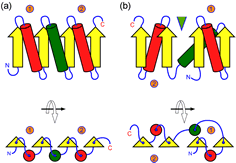
The figure at right (click on figure to show large version) shows two ways in which β-α-β units can combine to form a four-stranded parallel β sheet. In the figure, the β-α-β motifs are labeled 1 and 2, and the connecting helices are shown as green cylinders. Part (a) shows a sequential parallel sheet (the order of the strands is 1-2-3-4, from left to right, and all helices lie on the same face of the sheet. In part (b), the order of strands is 4-3-1-2, and the helices from motifs 1 and 2 lie on opposing faces of the sheet. In addition, in this case a crevice or cleft forms between motifs (indicated by the green triangle) at the sheet's C-terminal edge, which is often the location of binding sites for small-molecule ligands or substrates.
Methods for protein structure determination
Tertiary and quaternary structures are obtained experimentally by X-ray crystallography and multidimensional nuclear magnetic resonance (NMR) X-ray crystallography provides a high-resolution view of the structures of molecules. As applied to proteins, it typically reveals the locations of all non-hydrogen atoms that are well-localized in a tertiary structure to a resolution range of 3 - 1 Å. NMR provides a method of stucture determination complementary in a number of respects to crystallography. Hydrogen nuclei give rise to NMR signals, revealing the positions of hydrogen atoms. NMR provides a more dynamic picture of protein structures, which are in the case of NMR generally reported as ensembles of closely-related conformations.
X-ray crystallography of proteins: Seven easy steps to a structure
- Purification of protein in mg quantities
- Growth of crystals of “diffraction quality”
- Collect complete set of diffraction data
- Process data (“data reduction”)
- Solve the “phase problem”
- Build an initial model to fit the observed electron density
- Refinement: maximize agreement between model and data
Limtations of X-ray crystallography
- No crystal → no structure (e.g. membrane proteins are generally very difficult to crystallize)
- Parts of the molecule that are not fixed in conformation will not show up in the data
- Crystal structures are static pictures, while proteins are dynamic
- Effects of “crystal packing forces”: Is the crystal lattice a non-physiological state? (usually not considered a problem)
Evaluation of crystal structures
- What is the resolution of the data used to determine the structure?
- How closely does the model agree with the data (“R factor”)?
- Stereochemical quality of the model (e.g. are there φ, ψ outliers, wacky bond lengths, unfavorable bond angles, bad contacts?
- Is the structure supported by biochemical evidence?
Limitations of NMR
- Upper limit on size (in VVP4e, given as 100 kD)
- NMR structures are not “high-resolution"
- “Dynamic” features may be hard to distinguish from uncertainty in data
- The protein must be soluble to high concentration
Evaluation of NMR structures
- How “well-determined” is the structure? (The best NMR structures have 15 – 25 NOEs per residue.)
- How closely does the model agree with the data? (e.g. does the model predict observed NOEs?)
- Stereochemical quality of the model (e.g. are there φ, ψ outliers, wacky bond lengths, unfavorable bond angles, bad contacts?
- Is the structure supported by biochemical evidence?
Tertiary structures
General features of protein tertiary structure We can summarize much of what has been learned about protein structure in the form of some generalizations that describe features common to most if not all proteins.
- Proteins have well-packed, non-polar interiors, whereas polar and charged groups are accessible to the outside. Where polar groups occur in the interior (e.g. main chain carbonyls and amides), they tend to be paired.
- The interior of proteins are close-packed, i.e. there are no large cavities.
- The protein chains do not generally form "knots" (although see Taylor & Lin (2003).
- In comparing proteins, it is found that sequence (1° structure) similarity usually implies structural similarity. Note that there are numerous instances in which two proteins with little or no detectable sequence similarity nonetheless adopt very similar structures.
- Overall, there is very little strain in protein structures. In cases where local strain exists, the energetic cost is covered by numerous favorable interactions within the rest of the structure.
.gif)
.gif)
Quaternary structure
The top of the protein structure hierarchy is the description of how polypeptide chains with defined tertiary structures associate (noncovalently) to form stable complexes called multimers. The classic example illustrating the difference between tertiary and quaternary structure is the comparison between myoglobin (Mb) and hemoglobin (Hb).